
RESOLVING THE ANATOMY OF MATURE SCLERIFIED CONIFER SEED CONES: COMPLEMENTARITY AMONG THREE METHODS
DOI:
https://doi.org/10.5710/PEAPA.18.09.2022.425Keywords:
anatomy, conifer, microcomputed tomography, Taxodium, paraffin sectioning, seed cone, histologyAbstract
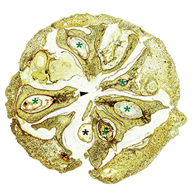
Phylogenetic studies of conifers that involve morphology are hindered by gaps in the anatomical characterization of seed cones, a direct result of difficulties encountered in sectioning cones in mature stages, which are often hardened due to sclerification. Here, we compare the resolving power of three methods—paraffin sectioning, petrographic thin-sectioning, and X-ray microcomputer tomography (micro-CT)—in documenting the morphology and anatomy of mature seed cones at different scales of detail. We use Taxodium as a case study, based on which we make recommendations on the complementarity of these methods, and we present a paraffin sectioning protocol for softening sclerified tissues. Paraffin sectioning, while providing high anatomical resolution, can only be used for small specimens, is labor-intensive, and hampered by hard tissues. Petrographic sectioning is fast and effective on larger specimens, but has low anatomical resolution and is limited to dry non-fleshy material. Micro-CT, if available, is fast, produces high resolution with no size limitations, and allows virtual sectioning and accurate 3D rendering; however, understanding of histology requires comparisons of CT images with results of the other methods. Although they overlap, each of the three methods provides unique insights on anatomy at different scales of detail. Thus, combining all three methods is ideal for producing high-quality data at all scales of anatomical and morphological detail.
References
Atkinson, B. A., Rothwell, G.A., & Stockey, R. A. 2014. Hughmillerites vancouverensis sp. Nov. and the Cretaceous diversication of Cupressaceae. American Journal of Botany, 101, 2136-2147.
Atkinson, B. A., Contreras D. L., Stockey, R. A., & Rothwell G. W. 2021. Ancient diversity and turnover of cunninghamioid conifers (Cupressaceae): two new genera from the Upper Cretaceous of Hokkaido, Japan. Botany, 99, 457-473.
Benedict, J. C. 2015. A new technique to prepare hard fruits and seeds for anatomical studies. Applications in Plant Sciences, 3, 1500075.
Escapa, I. H., & Catalano, S. A. 2013. Phylogenetic analysis of Araucariaceae: integrating molecules, morphology, and fossils. International Journal of Plant Sciences, 174, 1153-1170.
Herrera, F., Shi, G., Knopf, P., Leslie, A. B., Ichinnorov, N., Takahashi, M. Crane, P. R., & Herendeen, P. S. 2017. Cupressaceae conifers from the Early Cretaceous of Mongolia. International Journal of Plant Sciences, 178, 19-41.
Matsunaga, K.K.S., Herendeen, P.S., Herrera, F., Ichinnorov, N., Crane, P.R., Shi, G. 2021. Ovulate cones of Schizolepidopsis ediae sp. nov. provide insights into the evolution of Pinaceae. International Journal of Plant Sciences, 182, 490-507.
Rothwell, G. W., Stockey, R. A., Mapes, G., & Hilton, J. 2011. Structure and relationships of the Jurassic conifer seed cone Hughmillerites juddii gen. et comb. nov.: implications for the origin and evolution of Cupressaceae. Review of Palaeobotany and Palynology, 164, 45-59.
Smith, S.Y., Collinson, M.E., Simpson, D.A., Rudall, P.J., Marone, F., Stampanoni, M. 2009. Virtual taphonomy using synchrotron tomographic microscopy reveals cryptic features and internal structure of modern and fossil plants. Proceedings of the National Academy of Sciences USA, 106, 12013-12018.
Smith, S. Y., Stockey, R. A., Rothwell, G. W., & Little, S. A. 2016. A new species of Pityostrobus (Pinaceae) from the Cretaceous of California: moving towards understanding the Cretaceous radiation of Pinaceae. Journal of Systematic Palaeontology, 15, 69-81.
Stockey, R. A., Kvaček, J., Hill, R. S., Rothwell, G. W., & Kvaček, Z. 2005. The fossil record of Cupressaceae s. lat.. In A. Farjon (Ed.), A Monograph of Cupressaceae and Sciadopitys (pp. 54-68). Royal Botanic Garden Kew.
Additional Files
Published
Issue
Section
License
Copyright (c) 2022 Samar Riad El-Abdallah, Ashley Kammet, Kally Matsunaga, Selena Smith, Alexandru Tomescu

This work is licensed under a Creative Commons Attribution-NoDerivatives 4.0 International License.
Authors retain copyright and grant the journal right of first publication with the work simultaneously licensed under a Atribución/Reconocimiento 4.0 Internacional that allows others to share the work with an acknowledgement of the work's authorship and initial publication in this journal.